

The gut is home to a complex community of microorganisms, including bacteria, viruses and fungi. These microbes interact with the immune system, but the role of fungi—the gut mycobiome—in inflammatory bowel disease (IBD) is still not fully understood.
A new study published in the Journal of Crohn’s and Colitis examines how adaptive immune responses to fungal antigens appear in people with IBD. The researchers combined T cell repertoire sequencing with phage-immunoprecipitation sequencing (PhIP-Seq), a method that screens antibodies against a large collection of protein fragments.
The study found stronger signatures of fungal-specific T cells in people with Crohn’s disease than in those with ulcerative colitis. At the same time, the antibody analysis found relatively little shared reactivity to the yeast Saccharomyces cerevisiae. Together, the results offer a more detailed picture of antifungal immunity in IBD—and point to questions that future studies will need to address.
Looking at two parts of adaptive immunity
The adaptive immune system includes T cells and B cells, which help recognise and respond to specific targets.
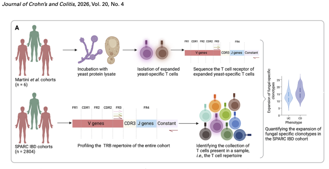
T cell receptor sequencing (TCR-Seq) identifies the receptor sequences carried by T cells in a sample. Groups of T cells with the same receptor sequence are called clonotypes. Some clonotypes have previously been linked to responses against particular fungal species, including Candida albicans, Candida tropicalis and S. cerevisiae. By tracking these clonotypes in a large IBD cohort, the researchers could compare their expansion across groups of people.
PhIP-Seq takes a different approach. It presents antibodies in a blood sample with a library of protein fragments displayed on bacteriophages. If antibodies bind to a fragment, researchers can identify the corresponding sequence. In this study, the team developed a library focused on S. cerevisiae proteins to investigate antibody responses to this yeast.
These methods provide complementary information: TCR-Seq tracks selected T cell signatures, while PhIP-Seq identifies protein fragments recognised by circulating antibodies.
A large cohort revealed stronger fungal T cell signatures in Crohn’s disease
For the T cell analysis, the researchers examined data from the SPARC IBD cohort, including 1,890 people with Crohn’s disease and 914 with ulcerative colitis. They found greater expansion of previously identified fungal-specific T cell receptor clonotypes in Crohn’s disease than in ulcerative colitis. This pattern included clonotypes associated with C. albicans, C. tropicalis and S. cerevisiae.
The researchers also examined a subset of T cell clonotypes previously characterised as Th1-type responses to fungi. Among people with Crohn’s disease, stronger S. cerevisiae-associated responses were linked to penetrating disease behaviour and perianal disease. These findings suggest that fungal-specific T cell signatures may be more prominent in some people with more complicated disease features.
The results show an association. They do not establish that fungal responses cause Crohn’s disease or make the disease more severe. The fungal-specific clonotypes used in the analysis had originally been identified in a small group of six donors, so the authors describe the population-level findings as requiring confirmation in larger and more diverse cohorts.
Connecting gut fungi with circulating T cells
The team next asked whether particular fungi detected in the gut were associated with particular T cell clonotypes in blood. They analysed paired gut mycobiome and T cell receptor data from 637 people with IBD.
This analysis identified 31 T cell clonotypes associated with 35 fungal or other microbial sequence variants that were prevalent enough to study. Most detected fungal sequence variants were found in relatively few people, making robust comparisons difficult.
These links are statistical associations: they show that a clonotype and a microbial sequence variant occurred together more often than expected under the analysis. They do not confirm that the T cells directly recognised that fungus. The authors also note that gut and blood samples were not always collected at the same time, which may have made associations harder to detect.
The limited number of robust links highlights a challenge in studying the gut mycobiome: fungal communities can vary substantially between individuals, while immune responses may also be concentrated in the gut rather than circulating in blood.
Antibody responses to yeast were limited in the PhIP-Seq analysis
To examine antibody responses, the researchers built a library containing more than 12,000 peptide fragments from 774 S. cerevisiae proteins. They tested blood serum from 100 people with Crohn’s disease and 60 healthy controls. The study groups included people with and without anti-S. cerevisiae antibodies, commonly abbreviated as ASCA.
Antibody reactivity to S. cerevisiae peptides was relatively low compared with responses to several viral and bacterial antigen groups included in the screening library. The analysis also found no single yeast peptide that was associated with ASCA status.
This does not mean that people with IBD have no immune response to fungi. PhIP-Seq detects antibodies that bind to protein fragments, but some fungal immune targets may not be captured by this approach. For example, ASCA are known to recognise carbohydrate-rich structures in fungal cell walls, which are not represented by the peptide fragments used in the study. Other targets may depend on protein shape or chemical modifications that peptide-display methods do not reproduce.
The researchers also found that ASCA positivity was associated with antibody reactivity to several bacterial flagellins, particularly those from members of the Clostridiales group. This points to broader patterns of immune recognition that may accompany ASCA status, rather than a response to one shared S. cerevisiae protein target.
What the findings mean—and what comes next
The study brings together large-scale T cell repertoire analysis, gut fungal profiling and a targeted antibody screen. Its findings suggest that fungal-specific T cell clonotypes are more expanded in Crohn’s disease than in ulcerative colitis, while shared antibody responses to the yeast peptides tested were limited.
The distinction between these results matters. Different parts of the immune system may recognise different targets, and those responses may not be equally visible in blood. A weak signal in a serum antibody screen does not rule out local immune activity in the gut, or responses to fungal structures that the assay was not designed to detect.
Future work could broaden the range of fungal species and antigens studied, include targets such as cell-wall carbohydrates and virulence-associated proteins, and examine immune cells and antibodies directly in gut tissue. Larger and more genetically diverse cohorts could also help researchers identify fungal-specific T cell responses across a wider range of HLA backgrounds.
By combining complementary immune-profiling methods, this research adds detail to the picture of host–microbiome interactions in IBD. It also shows why understanding antifungal immunity will require methods that capture the diversity of fungal targets and the different places where immune responses take place.
Publication
Aya K.H. Mahdy, Hesham ElAbd, Melanie Prinzensteiner, Hannah Jebens, Kostas Sivickis, Petra Bacher, Thomas Vogl, Mathilde Poyet and Andre Franke. “Mapping adaptive immune responses toward fungal antigens in inflammatory bowel disease using T cell repertoire sequencing and phage-immunoprecipitation sequencing.” Journal of Crohn’s and Colitis 20, issue 4 (2026), jjag015. Read the publication.